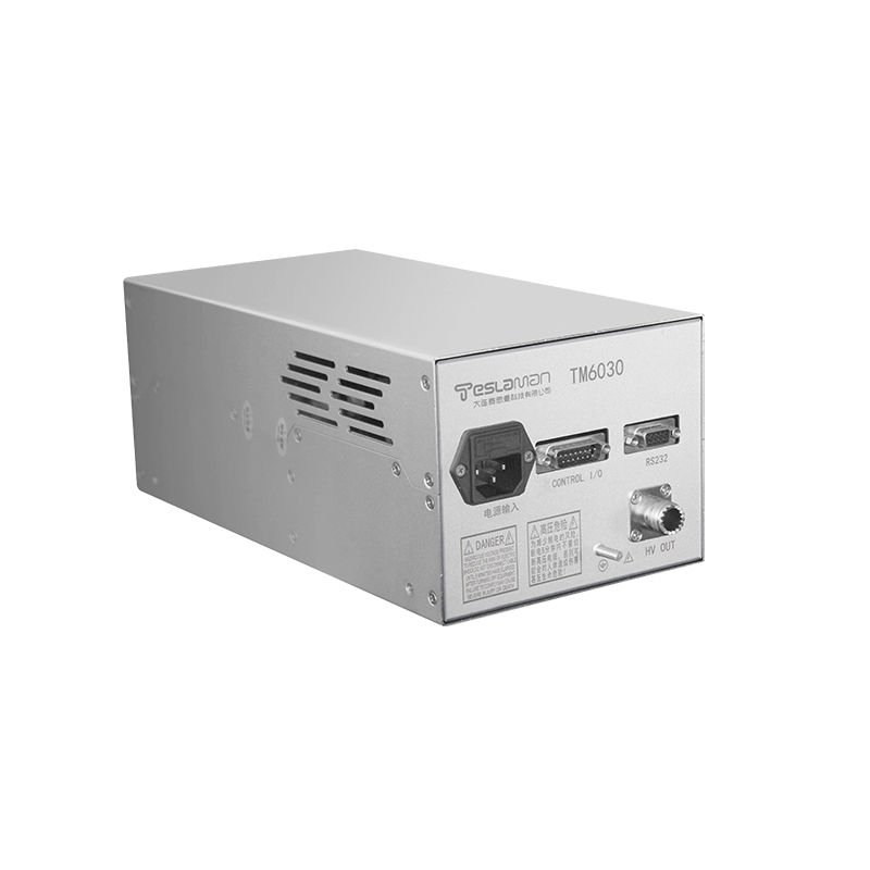
Optimization of High-Voltage Power Supply for Mammographic Imaging
As the core component of mammography systems, the performance of high-voltage power supplies directly impacts image quality and diagnostic accuracy. Recent advancements in high-frequency inversion, power factor correction, target material innovation, and system integration have significantly optimized mammographic imaging in precision, dose control, and spatial adaptability.
1. High-Frequency Inversion and Ripple Control
Early line-frequency power supplies suffered from high output ripple (>10%), causing image artifacts and blurred details. Modern high-frequency inversion technology elevates load inversion frequency to 80kHz (idle up to 100kHz), reducing ripple coefficients to <1% and significantly enhancing image clarity. High-frequency transformers with high-permeability materials achieve >95% efficiency. Coupled with electronic rectification modules (e.g., Schottky diodes and 4700μF filter capacitors), output voltage fluctuation is controlled within 0.5%, ensuring X-ray beam stability and reducing missed diagnoses of microcalcifications.
2. Power Factor Correction (PFC) and Output Stability
PFC technology corrects power noise by dynamically regulating input current/voltage waveforms and frequency, increasing the power factor to >0.99. During rectification-inversion, PFC compresses voltage/current fluctuations to <5%, yielding near-zero ripple in DC output. This homogenizes X-ray energy distribution and boosts image contrast by ~30%. Experiments show spatial resolution can reach 0.133mm—sufficient to resolve early cancerous lesions.
3. Dual-Target Technology and Dose Optimization
For breast soft-tissue imaging, power supplies require specialized target materials. Traditional molybdenum (Mo) targets exhibit poor heat resistance, accelerating tube degradation. The dual-target design (Mo base + rhodium (Rh) ring) enables dynamic electron beam switching:
Rh mode: Generates high-energy X-rays at 28-35kV for dense breasts (thickness ≥5cm), reducing dose by 15%;
Mo mode: Optimized for 20-28kV imaging, enhancing contrast between adipose and glandular tissues.
Studies confirm that Mo/Rh combinations reduce mean glandular dose (MGD) by 20% versus Mo/Mo for 4cm breasts while improving contrast-to-noise ratio (CNR) by 18%.
4. Integrated Tube Head Design
Miniaturization and cable-free integration represent breakthroughs. Direct coupling of high-voltage generators and X-ray tubes eliminates high-voltage cables, shrinking tube head length to <30cm. This triple optimization offers:
Enhanced safety: Prevents cable aging risks, with leakage current <10mA;
Space adaptability: Fits compact rooms in community clinics;
Energy efficiency: Reduces power transmission loss by 40% and cooling demands.
5. Dynamic Parameter Tuning Strategy
Based on Chinese women’s breast characteristics (glandular content: 20%-50%), voltage (kV), target/filter combinations, and exposure (mAs) require dynamic synergy:
Figure-of-merit (FOM) guidance: FOM=CNR²/MGD peaks at 29kV with Mo/Rh for 4cm phantoms;
Real-time control: Embedded systems (e.g., CompactRIO) regulate focus electrode voltage with <0.5% error, increasing electron transmission from <20% to >50%.
Conclusion
High-voltage power supply optimization drives mammographic imaging advancements. Through high-frequency/low-ripple output, dual-target dose control, miniaturization, and intelligent parameter tuning, submillimeter resolution is achieved while reducing radiation risks. Future AI-driven adaptive voltage regulation and semiconductor materials will further enhance screening precision and safety.


