


Relationship Between Kilovolt and Milliampere Three-dimensional Adjustment of Cone-beam CT High Voltage Power Supply and Imaging Quality
Cone-beam computed tomography provides three-dimensional imaging for medical and industrial applications. The X-ray tube voltage and current determine the imaging characteristics. Three-dimensional adjustment of kilovolt and milliampere enables optimization for different applications. Understanding the relationship between electrical parameters and imaging quality enables optimal image acquisition.
Cone-beam CT fundamentals involve X-ray projection and reconstruction. An X-ray source rotates around the object. A flat panel detector captures projection images. Multiple projections are acquired from different angles. Reconstruction algorithms create a three-dimensional volume. The image quality depends on the acquisition parameters.
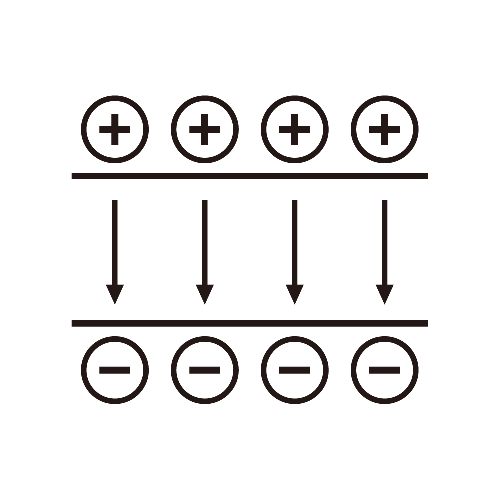
X-ray tube voltage effects on imaging are significant. The voltage determines the X-ray energy spectrum. Higher voltage produces higher energy X-rays. Higher energy X-rays penetrate denser materials. The voltage affects the contrast. The voltage must be appropriate for the object.
X-ray tube current effects on imaging are important. The current determines the X-ray flux. Higher current produces more photons. More photons reduce quantum noise. The current affects the signal-to-noise ratio. The current must be adequate for the application.
Exposure time effects on imaging quality are significant. Longer exposure increases the total dose. Higher dose reduces noise. However, longer exposure increases motion artifacts. The exposure must be optimized. The optimization must balance multiple factors.
Three-dimensional parameter space requires systematic exploration. The voltage, current, and time form a parameter space. Each point in the space produces different image quality. The optimal point depends on the application. The exploration must be systematic. The exploration must be practical.
Contrast-to-noise ratio is a key quality metric. The contrast depends on the voltage. The noise depends on the current and time. The ratio determines the detectability. The ratio must be optimized. The optimization must consider the application.
Spatial resolution depends on multiple factors. The focal spot size affects the resolution. The detector resolution affects the resolution. The geometry affects the resolution. The voltage and current affect the focal spot. The resolution must meet the requirements.
Artifacts can degrade the image quality. Beam hardening causes cupping artifacts. Scatter reduces contrast. Motion causes blurring. The parameters affect the artifacts. The artifacts must be minimized.
Dose considerations are important for medical applications. The dose must be minimized while maintaining quality. The dose depends on the voltage, current, and time. The dose must be appropriate for the diagnostic task. The optimization must consider the dose.
Application-specific optimization is necessary. Different body parts require different parameters. Different materials require different settings. The optimization must be specific to the application. The optimization must be validated. The optimization must be practical.
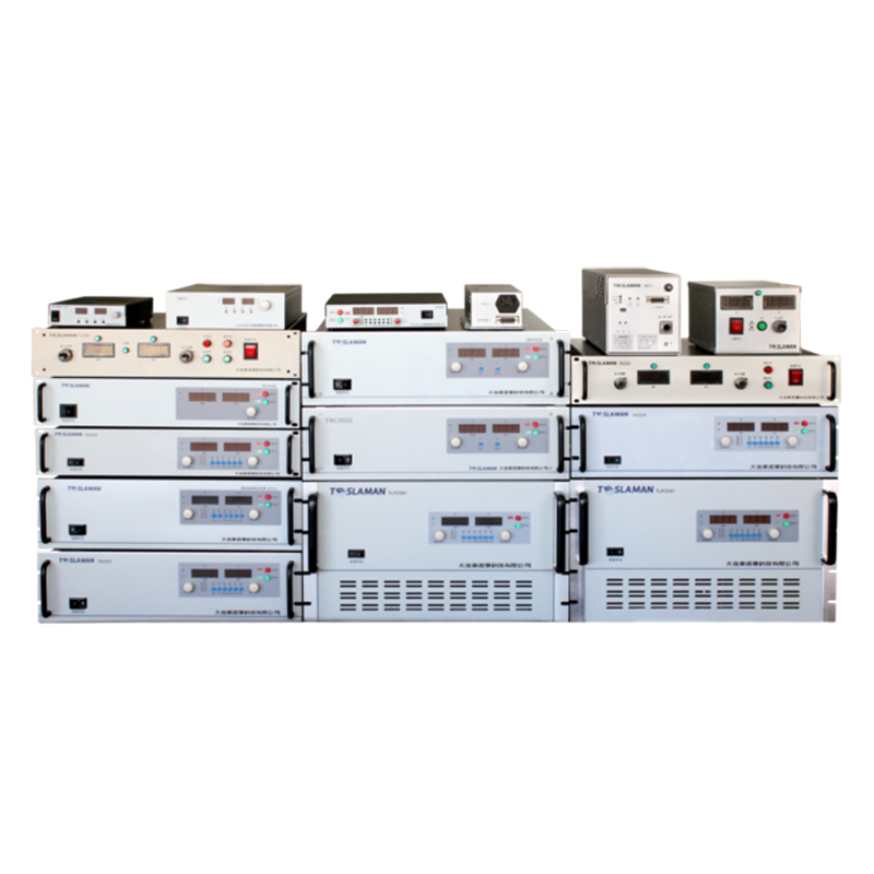
Calibration of the imaging system is essential. The voltage and current must be accurate. The calibration must be traceable. The calibration must be maintained. The calibration affects the image quality. The calibration must be documented.
Quality control ensures consistent imaging. Regular testing verifies the performance. The quality control must be comprehensive. The documentation must support the quality. The quality control must meet standards.
Research methodology for parameter optimization requires systematic approach. Phantom studies enable controlled experiments. Clinical studies validate the optimization. Statistical analysis identifies significant effects. The methodology must be rigorous. The research must be validated.