


Electric Field Parameter Window Exploration for High Voltage Pulsed Electric Field Induced Tumor Cell Apoptosis
High voltage pulsed electric field therapy has emerged as a promising modality for cancer treatment, offering the potential to induce tumor cell death through non-thermal mechanisms that avoid the collateral damage associated with conventional treatments. The technique employs brief high voltage pulses to permeabilize cell membranes and trigger intracellular signaling cascades that lead to apoptosis. Identifying the optimal electric field parameter window that maximizes tumor cell killing while sparing normal tissue requires systematic exploration of the complex relationships between pulse characteristics and biological responses.
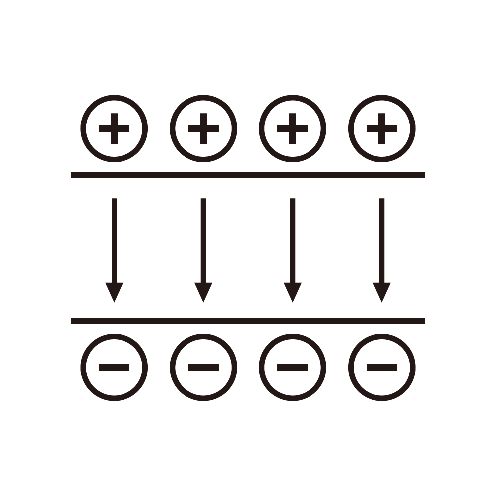
The fundamental mechanism of pulsed electric field effects on cells involves electroporation of the cell membrane. When an electric field of sufficient strength is applied, the transmembrane potential increases until it exceeds a critical threshold, causing structural rearrangement of the lipid bilayer and formation of pores. These pores enable enhanced transport of molecules across the membrane, potentially triggering intracellular signaling pathways that lead to programmed cell death.
Apoptosis represents a form of programmed cell death characterized by specific morphological and biochemical changes. Cells undergoing apoptosis exhibit membrane blebbing, nuclear condensation, DNA fragmentation, and formation of apoptotic bodies. This controlled death process avoids the inflammatory response associated with necrosis, potentially offering therapeutic advantages. Pulsed electric fields can induce apoptosis through multiple mechanisms involving membrane permeabilization, calcium influx, and intracellular signaling activation.
The electric field strength represents the primary parameter determining the extent of membrane effects. Field strengths below the electroporation threshold cause minimal membrane perturbation. Field strengths above the threshold cause reversible or irreversible permeabilization depending on the magnitude and duration. The optimal field strength for apoptosis induction lies in a range that causes sufficient membrane perturbation to trigger death pathways without causing immediate necrosis.
Pulse duration significantly influences the nature of membrane effects and cellular responses. Microsecond pulses can cause reversible electroporation with membrane recovery after pulse cessation. Nanosecond pulses can affect intracellular structures in addition to the plasma membrane. Millisecond pulses can cause more extensive membrane damage. The pulse duration must be optimized for the desired balance of membrane and intracellular effects.
Pulse number affects the cumulative membrane perturbation and cellular stress. Single pulses may cause transient effects that cells can recover from. Multiple pulses can cause cumulative stress that triggers apoptosis. Excessive pulse numbers may cause immediate necrosis rather than apoptosis. The pulse number must be optimized to achieve apoptosis without causing necrosis.
Pulse repetition frequency influences the time available for cellular recovery between pulses. Lower frequencies allow more complete recovery, potentially requiring more pulses to achieve cumulative effects. Higher frequencies may cause cumulative stress more efficiently but could also favor necrosis. The repetition frequency must be coordinated with pulse number and duration for optimal effects.
Pulse waveform shape affects the membrane charging dynamics and the resulting electroporation characteristics. Square pulses provide constant field strength throughout the pulse duration. Exponential pulses provide decaying field strength that may cause different membrane effects. Bipolar pulses may reduce electrolysis effects and provide symmetric membrane perturbation. The waveform shape must be selected for appropriate electroporation behavior.
Cell type characteristics significantly influence the optimal parameter window. Different cell types have different membrane compositions, sizes, and geometries that affect electroporation thresholds. Tumor cells may have different characteristics than normal cells, potentially enabling selective effects. The parameter optimization must account for the specific cell types involved in the treatment.
Tissue characteristics affect the electric field distribution and the resulting cellular exposure. Tissue conductivity and permittivity determine the field penetration and distribution. Tissue heterogeneity causes non-uniform field exposure for cells at different locations. The treatment planning must account for tissue characteristics to ensure appropriate field delivery to target cells.
Temperature effects on electroporation and apoptosis influence the parameter optimization. Temperature affects membrane fluidity, electroporation threshold, and cellular stress responses. Elevated temperatures can enhance electroporation effects and potentially synergize with electric field treatment. The temperature conditions must be considered in parameter selection.
In vivo considerations for parameter optimization include the challenges of field delivery in living tissue. Electrode configuration determines the field distribution in the treatment volume. Tissue movement and physiological changes can affect field delivery during treatment. The in vivo parameter window may differ from in vitro observations due to tissue effects.
Safety considerations for clinical application require identification of parameter ranges that avoid harmful effects. Excessive field strength or pulse energy can cause tissue damage beyond therapeutic effects. Electrical stimulation of nerves and muscles can cause discomfort or functional disruption. Thermal effects from pulse energy dissipation must be controlled to avoid hyperthermia. The safety window must be established alongside the therapeutic window.
Treatment efficacy assessment requires appropriate metrics for evaluating apoptosis induction. Cell viability assays quantify surviving cells after treatment. Apoptosis markers identify cells undergoing programmed death pathways. Tumor growth measurements evaluate therapeutic effects in animal models. The efficacy metrics must capture the relevant therapeutic outcomes.
Parameter window mapping systematically explores the relationship between pulse parameters and biological effects. Multi-dimensional parameter sweeps test various combinations of field strength, duration, number, and frequency. Response surface modeling characterizes the parameter-effect relationships across the explored space. Optimization algorithms identify parameter combinations that maximize therapeutic effects.
Clinical translation of optimized parameters requires validation in appropriate models. Animal studies verify that parameters optimized in vitro achieve therapeutic effects in vivo. Safety studies confirm that parameters avoid harmful effects in clinical-relevant conditions. Clinical trials evaluate therapeutic efficacy and safety in human patients. The translation process must bridge laboratory findings to clinical practice.
Continued research in pulsed electric field cancer therapy drives ongoing exploration of parameter windows. Better understanding of electroporation and apoptosis mechanisms enables more targeted parameter selection. Advanced pulse generation technology provides improved control over treatment parameters. Integration with imaging and monitoring enables adaptive parameter adjustment during treatment. These developments continue to advance the potential for pulsed electric field therapy in cancer treatment.