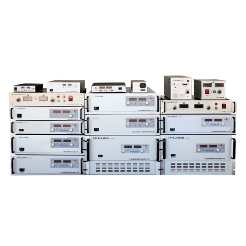


Application and Technical Challenges of High-Voltage Power Supplies in Nuclear Medicine
Nuclear medicine, a critical branch of modern medicine, relies on radionuclide tracing technology to achieve precise diagnosis and treatment of human physiological functions and diseases. As one of the core components of nuclear medicine equipment, high-voltage power supplies (HVPS) directly influence imaging quality, radiation safety, and therapeutic efficacy. This article discusses the key application scenarios of HVPS in nuclear medicine from a professional perspective and analyzes their technical challenges and development trends.
1. Core Application Scenarios of HVPS in Nuclear Medicine
1. Radionuclide Production and Labeling
Nuclear medicine imaging (e.g., PET, SPECT) depends on the preparation of short-half-life radionuclides (e.g., ¹⁸F, ⁹⁹mTc). HVPS are used in cyclotrons to accelerate charged particles (e.g., protons, deuterons) for bombarding target materials in nuclear reactions. For instance, in ¹⁸F production, HVPS must provide stable megavolt-level voltages with energy accuracy better than ±0.1% to control reaction yields and impurity formation. Additionally, in radiopharmaceutical labeling, HVPS-driven electrospray ionization enables precise preparation of nanoscale radioactive probes, enhancing labeling efficiency and stability.
2. Signal Amplification in Nuclear Imaging Devices
Scintillation detectors (e.g., NaI(Tl), LSO crystals) are core components of nuclear imaging, and their weak current signals (picometer level) require amplification by photomultiplier tubes (PMTs) or silicon photomultipliers (SiPMs) driven by HVPS. In PET systems, for example, HVPS must supply stable bias voltages (800–1500V) to PMTs with noise levels below 10mV RMS to avoid signal distortion. Dynamically adjustable HV modules can adapt to the gain requirements of different crystal materials, optimizing imaging contrast and spatial resolution (e.g., reducing PET spatial resolution from 4mm to below 2mm).
3. Precise Dose Control in Radiotherapy
In nuclear medicine therapy (e.g., β-ray applicator treatment, targeted radiotherapy), HVPS drive electron accelerators to generate high-energy electron beams. For skin cancer treatment, HVPS must adjust electron energy to 5–20MeV to ensure precise dose deposition within tumor depths (0.5–5mm) while protecting normal tissues. Moreover, the fast response (switching time <10μs) of HVPS enables pulsed radiotherapy, which, combined with real-time dose monitoring, limits dose errors to ±3%.
2. Technical Challenges Facing HVPS
1. Stability and Reliability in Extreme Environments
Nuclear medicine equipment often operates in high-humidity, high-magnetic-field, or high-radiation environments, where the insulating materials of HVPS are prone to radiation damage (e.g., polymer degradation from ionizing radiation), leading to partial discharge or breakdown. In proton therapy rooms, for example, long-term neutron radiation may change the dielectric constant of HV modules by over 10%, causing output voltage drift. Developments in radiation-resistant insulating materials (e.g., ceramic composites, fluoropolymers) and optimized thermal management (e.g., microchannel liquid cooling) are needed to control temperature fluctuations within ±1℃.
2. Synergetic Control Requirements for Multimodal Imaging
With the development of nuclear medicine toward multimodal fusion (e.g., PET/MRI, SPECT/CT), HVPS must be compatible with magnetic field and radiofrequency systems. Electromagnetic interference (EMI) from traditional HVPS may disrupt MRI gradient fields (e.g., introducing >50nT noise), causing image artifacts. Magnetic shielding (e.g., multi-layer permalloy encapsulation) and high-frequency soft-switching topologies (e.g., LLC resonant converters) are required to suppress EMI noise below 100dBμV while achieving power efficiency >95%.
3. Miniaturization and Low-Power Design Bottlenecks
Portable nuclear medicine devices (e.g., mobile PET scanners) require HVPS to be reduced to 1/5 of traditional modules’ volume, with power density exceeding 500W/in³. Conventional power supplies based on line-frequency transformers are inadequate; planar magnetic core technology (e.g., thin-film transformers) and wide-bandgap semiconductors (e.g., SiC MOSFETs) are needed to increase switching frequency to the MHz range, while 3D stacking packaging reduces volume. Standby power must also be lowered from watts to milliwatts to meet battery life demands in field or emergency scenarios.
4. Radiation Safety and Electromagnetic Compatibility Design
HV components of power supplies may act as secondary radiation sources, requiring lead shielding (>5mm thickness) to attenuate X-rays to <1μSv/h. During equipment integration, HV cable layouts must be optimized to avoid electromagnetic coupling with signal cables (e.g., crosstalk rejection ratio >60dB), preventing increased data bit error rates in imaging.
3. Technical Development Trends and Prospects
Future HVPS development in nuclear medicine will focus on intelligence, integration, and sustainability:
Intelligence: Adaptive control algorithms (e.g., fuzzy PID, neural networks) will enable dynamic optimization of HV parameters, such as automatically adjusting PMT bias voltages in PET detectors based on patient body size to enhance imaging signal-to-noise ratio.
Integration: System-in-package (SiP) technology will integrate HVPS with signal processing circuits into a single module, reducing signal transmission paths and parasitic parameter effects.
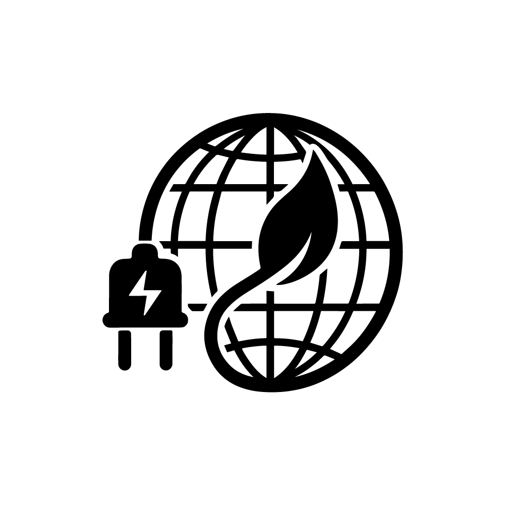
Sustainability: Development of renewable energy-compatible HVPS systems (e.g., solar-powered radionuclide production) will reduce carbon footprints, while recycling rates for obsolete power supplies aim to exceed 90%.
In summary, breakthroughs in HVPS technology will continue to drive nuclear medicine toward precision and portability, providing safer and more efficient technical support for disease diagnosis and treatment.