


Shear Wave Excitation Control of High Voltage Transmission Power Supply for Medical Ultrasound Elastography
Ultrasound elastography has emerged as a valuable diagnostic technique for assessing tissue stiffness, providing information complementary to conventional B mode imaging that can aid in detection and characterization of tumors, liver fibrosis, and other pathological conditions. The technique relies on generating shear waves within tissue and tracking their propagation to infer mechanical properties. High voltage transmission power supplies drive the ultrasound transducer elements that produce the acoustic radiation force impulses responsible for shear wave generation, with precise control of the excitation parameters essential for accurate and reproducible elastography measurements.
Shear wave elastography operates on the principle that the shear wave propagation speed in tissue depends on the underlying tissue stiffness. Acoustic radiation force impulse pushing beams generate localized tissue displacement that launches shear waves propagating away from the push location. By measuring the time required for the shear wave to travel between two tracking locations, the shear wave speed can be calculated and related to tissue stiffness through appropriate mechanical models. The quality of the shear wave and the accuracy of the speed measurement depend critically on the characteristics of the pushing beam.
The acoustic radiation force magnitude depends on the acoustic intensity of the pushing beam and the acoustic properties of the tissue. Higher intensity beams produce larger displacements and stronger shear waves, improving the signal to noise ratio for wave tracking. However, acoustic intensity is limited by safety considerations including mechanical index and thermal index constraints that prevent tissue damage from excessive acoustic exposure. The high voltage power supply must provide sufficient output to achieve the desired pushing beam intensity while enabling precise control to stay within safety limits.
Pulsed excitation is used for acoustic radiation force impulse generation, with the pulse duration affecting both the displacement magnitude and the spatial extent of the push. Longer pulses deliver more energy and produce larger displacements, but also broaden the push region in the axial direction, reducing the spatial resolution of the elastography measurement. Typical pulse durations for shear wave elastography range from tens to hundreds of microseconds, requiring the power supply to maintain stable output throughout the pulse duration and to switch off rapidly at the pulse end.
The voltage waveform applied to the transducer elements determines the acoustic pressure waveform and thus the characteristics of the pushing beam. Conventional excitation uses rectangular pulses that produce broadband acoustic signals with energy distributed across a wide frequency range. Toneburst excitation at the transducer resonant frequency concentrates the acoustic energy in a narrower bandwidth, improving efficiency and reducing unwanted frequency components. The power supply output characteristics must support the desired waveform shape with adequate fidelity.
Channel to channel matching in multi element transducer arrays affects the uniformity of the pushing beam and the resulting shear wave. Variations in the high voltage output across channels create nonuniform acoustic output that distorts the beam profile and produces asymmetric shear wave patterns. The power supply design must ensure that all channels receive matched excitation, with tight tolerances on output voltage and timing across the array. Calibration procedures verify and adjust channel matching to maintain beam quality.
The timing relationship between the pushing pulse and the tracking pulses used to detect shear wave motion must be precisely controlled. The tracking pulses are transmitted at varying time delays after the push to sample the tissue displacement at different times during shear wave propagation. The delay resolution determines the temporal sampling of the shear wave and thus the accuracy of speed measurement. High resolution timing control in the power supply and system sequencing enables fine sampling of the shear wave passage.
Safety monitoring systems track the acoustic output and thermal exposure to ensure compliance with regulatory limits and safe clinical practice. The high voltage power supply output is monitored and accumulated to track the mechanical and thermal exposure over the examination. Automatic shutdown systems disable the high voltage output if safety limits are approached, preventing excessive exposure. These safety systems must be reliable and fail safe, as failure to limit acoustic output could result in patient injury.
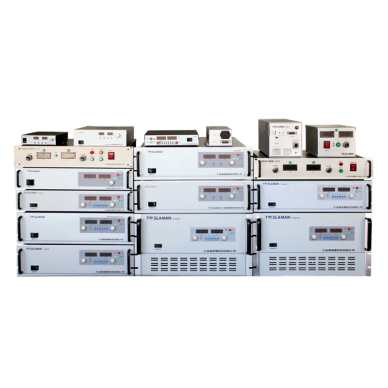
The transducer type and configuration influence the power supply requirements for shear wave elastography. Single element transducers require simpler power supply architectures with a single high voltage channel. Array transducers require multichannel power supplies with independent control of each element or group of elements. The aperture configuration for pushing and tracking beams affects the number of active channels and the power delivery requirements. Power supply design must accommodate the specific transducer characteristics and the beamforming approaches used for elastography.
Advanced elastography techniques place additional demands on the high voltage power supply. Supersonic shear imaging uses multiple pushing pulses in rapid succession to create a shear wave source that effectively moves faster than the shear wave speed, generating a high amplitude Mach cone shear wave. This technique requires multiple high voltage pulses with precise timing intervals, challenging the power supply repetition rate and timing precision. Comb push elastography uses simultaneous multiple push beams to interrogate multiple locations, requiring multiple channel excitation with controlled phase relationships.