


Beamforming and Focusing Accuracy Control of High Voltage Transmitter Power Supply for Ultrasound Imaging
Medical ultrasound imaging uses phased arrays of transducer elements to generate and receive ultrasound waves for visualizing internal anatomy. The beamforming process controls the timing and amplitude of signals to each element, creating a focused beam that scans through the tissue. The high voltage transmitter power supply that drives the transducer elements must provide precise voltage and timing control for accurate beamforming and focusing.
Ultrasound transducer arrays consist of many piezoelectric elements that convert electrical signals to acoustic waves and vice versa. Typical arrays have 64 to 256 elements arranged in linear, curved, or two dimensional configurations. Each element is driven by a high voltage pulse that causes the piezoelectric material to vibrate, producing an ultrasound wave. The timing of pulses to different elements controls the beam direction and focus.
Beamforming for transmission applies delays to the pulses for each element so that the acoustic waves from all elements arrive at a focal point simultaneously. The delays compensate for the different path lengths from each element to the focal point. Elements farther from the focal point receive pulses earlier, so their waves travel longer and arrive simultaneously with waves from closer elements. The delay pattern determines the beam direction and the focal depth.
Focusing accuracy depends on the precision of the delay timing. The ultrasound wavelength in tissue is approximately 0.5 millimeters for typical diagnostic frequencies. The timing precision must be a fraction of the wave period to achieve accurate focusing. For 5 MHz ultrasound, the period is 200 nanoseconds, requiring timing precision of tens of nanoseconds for accurate beamforming.
The high voltage transmitter power supply provides the excitation pulses to the transducer elements. The pulse voltage determines the acoustic amplitude, affecting the imaging penetration and resolution. Higher voltages produce stronger acoustic waves that penetrate deeper but may cause increased tissue heating. The voltage must be controlled precisely for consistent imaging performance.
Pulse waveform characteristics affect the acoustic output. The pulse duration determines the frequency content and the axial resolution. Shorter pulses provide better axial resolution but may have broader frequency bandwidth. The pulse rise time affects the high frequency content. The pulse shape can be optimized for specific imaging applications.
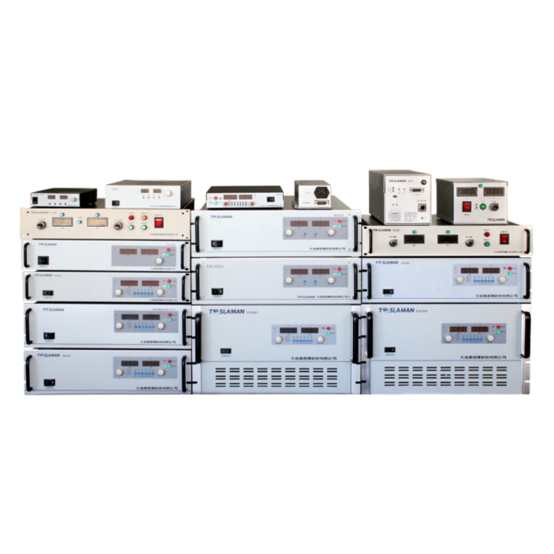
Multi channel transmitter design provides independent control of each transducer element. Each channel has its own high voltage driver with programmable delay and amplitude control. The channels operate in parallel to generate the simultaneous pulses for beamforming. The channel count matches the transducer array configuration.
Channel synchronization ensures that all channels fire with the correct timing relationships. A master clock provides the timing reference for all channels. Delay generators produce the individual channel triggers with programmable delays from the master clock. The synchronization must maintain timing accuracy across all channels despite variations in component characteristics.
Voltage consistency across channels affects the beam uniformity. Variations in pulse voltage between channels cause variations in acoustic amplitude, affecting the beam profile and the image uniformity. The transmitter design must provide consistent voltage across all channels. Calibration can compensate for any systematic variations.
Dynamic focusing changes the focal depth during imaging to maintain focus throughout the imaging depth range. The delays are updated as the beam scans through different depths, keeping the focal point at the current imaging depth. Dynamic focusing requires rapid delay updates, potentially for each transmit event. The transmitter must support the required delay update rate.
Aperture control varies the number of active elements to optimize the beam characteristics. Larger apertures provide better lateral resolution but have longer near field lengths. Smaller apertures have shorter near field but wider beams. The aperture may be varied with depth to optimize the resolution throughout the image. The transmitter must enable flexible aperture control through channel selection.
Harmonic imaging uses the nonlinear propagation of ultrasound to generate harmonic frequencies that can improve image quality. The transmitter pulses are designed to optimize harmonic generation, typically using specific pulse shapes or amplitudes. The high voltage supply must provide the appropriate pulse characteristics for harmonic imaging modes.