














Microfluidic Chip Cell Electrofusion High Voltage Pulse Power Supply Microelectrode Array Design and Optimization
The integration of high voltage pulse power supply systems with microfluidic chip platforms represents a significant advancement in cell electrofusion technology. Cell electrofusion, a process that enables the merging of two or more cells through electrical stimulation, has become an indispensable tool in biomedical research, somatic cell nuclear transfer, and hybridoma production for monoclonal antibody generation. The microfluidic chip approach offers precise control over cell pairing, reduced sample consumption, and improved fusion efficiency compared to conventional bulk electrofusion methods.
The fundamental principle underlying cell electrofusion involves the application of high voltage pulses to create temporary pores in cell membranes, facilitating cytoplasmic exchange between adjacent cells. The pulse power supply must generate electric field strengths sufficient to induce reversible electroporation without causing irreversible membrane damage. Typical field requirements range from 1 to 10 kV/cm, with pulse durations spanning microseconds to milliseconds depending on cell type and desired fusion outcomes.
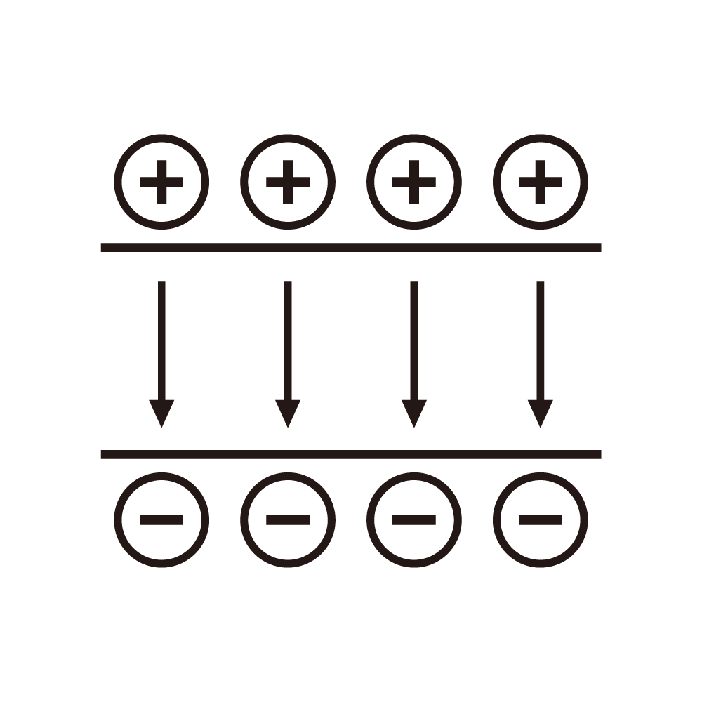
Microelectrode array design constitutes the critical interface between the high voltage power supply and the biological sample. Several electrode geometries have been investigated, including parallel plate configurations, interdigitated arrays, and three-dimensional electrode structures. Parallel plate electrodes offer uniform electric field distribution but limited cell positioning control. Interdigitated arrays enable selective addressing of individual electrode pairs but introduce field non-uniformities near electrode edges. Three-dimensional electrode structures, fabricated using microelectromechanical systems technology, provide enhanced field concentration within microchannels while maintaining cell viability.
The optimization of electrode dimensions requires careful consideration of multiple parameters. Electrode spacing directly influences the required voltage amplitude for a given electric field strength. Narrower gaps reduce voltage requirements but increase fabrication complexity and may restrict cell passage through microchannels. Typical electrode gaps in microfluidic electrofusion devices range from 20 to 200 micrometers, with the optimal dimension determined by cell size, channel geometry, and power supply constraints. Electrode width affects the uniformity of the electric field across the fusion zone, with wider electrodes providing more consistent field distribution.
Material selection for microelectrode fabrication involves trade-offs between electrical conductivity, chemical stability, and biocompatibility. Gold remains the most commonly employed electrode material due to its excellent conductivity and resistance to oxidation. However, gold electrodes may require adhesion layers such as chromium or titanium when deposited on glass or silicon substrates. Platinum and platinum-iridium alloys offer superior electrochemical stability during pulse operation but at higher material costs. Indium tin oxide provides optical transparency for microscopy-based observation but exhibits lower conductivity than metallic alternatives.
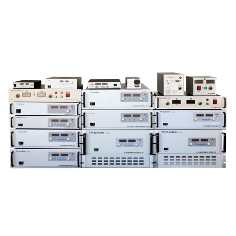
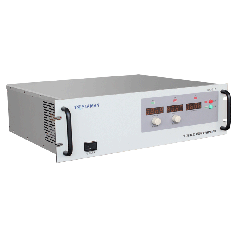
The high voltage pulse power supply design for microfluidic cell electrofusion must address several technical challenges. Pulse amplitude requirements typically range from 10 to 100 volts for microelectrode configurations, substantially lower than bulk electrofusion systems due to reduced electrode spacing. Rise time and fall time characteristics influence pore formation dynamics and should be minimized to achieve sharp field transitions. Pulse repetition rate capability enables multiple fusion attempts on cell pairs that fail to fuse during initial pulse application.
Capacitive discharge circuits represent the predominant architecture for microfluidic electrofusion pulse generation. Energy storage in capacitor banks enables rapid energy release during pulse formation, while switching elements such as insulated gate bipolar transistors or metal oxide semiconductor field effect transistors control pulse timing and duration. The capacitor bank voltage determines the maximum pulse amplitude, while capacitance value affects pulse shaping during resistive and capacitive loading from the electrode-cell system.
Load impedance characterization constitutes an essential step in power supply design optimization. The electrical impedance of microfluidic electrofusion devices varies with buffer conductivity, electrode geometry, and cell density. Higher conductivity buffers reduce impedance but increase joule heating during pulse application. Temperature rise calculations must ensure that thermal effects remain within acceptable limits for cell viability. Real-time impedance monitoring during the electrofusion process enables adaptive pulse parameter adjustment for varying biological samples.
Field uniformity across the fusion zone directly impacts fusion yield and cell viability. Finite element analysis provides valuable insights into electric field distribution within complex electrode geometries. Simulation results guide electrode design modifications to minimize field hotspots that cause localized membrane damage while ensuring adequate field strength at cell contact regions. Dielectrophoretic cell alignment prior to pulse application benefits from field gradient regions near electrode edges, creating an additional design consideration for electrode shape optimization.
Pulse parameter optimization studies have established relationships between electrical parameters and fusion outcomes. Square wave pulses offer well-defined duration and amplitude control, while exponential decay pulses from capacitive discharge provide simpler circuit implementation. Optimal pulse duration typically ranges from 10 to 100 microseconds for most mammalian cell types, with shorter pulses requiring higher field strengths to achieve equivalent membrane permeabilization. Multiple pulse applications with controlled interpulse intervals can enhance fusion efficiency without cumulative damage effects.
Thermal management during pulse operation requires careful attention in microfluidic systems. The high surface-to-volume ratio in microchannels provides enhanced heat dissipation compared to bulk systems, but localized heating near electrode surfaces may still affect cell viability. Integration of temperature sensors within the chip enables real-time thermal monitoring during pulse sequences. Pulse protocol design incorporating cooling intervals between pulse bursts prevents excessive temperature rise during extended fusion operations.
The integration of cell positioning mechanisms with electrofusion systems enables automated fusion protocols. Dielectrophoretic trapping using alternating current fields brings cells into close contact prior to high voltage pulse application. Optical tweezers provide non-contact cell manipulation but require complex optical setups. Hydrodynamic focusing through microchannel design guides cells into fusion zones with high throughput. The selection of cell positioning method influences electrode design and power supply requirements for the overall system.
Quality control and reproducibility in microfluidic electrofusion demand standardized characterization procedures. Electric field mapping using electrooptic sensors or electrolyte solutions provides quantitative assessment of field uniformity. Fusion efficiency measurement requires consistent cell counting methods before and after pulse application. Viability assays distinguish fused cells from damaged cells that have undergone irreversible membrane rupture. Statistical analysis of fusion outcomes across multiple experimental runs establishes protocol reliability.