Low-Voltage Cathodoluminescence Excitation in Scanning Electron Microscopy: The Critical Role of High-Voltage Stability
Cathodoluminescence, the emission of light from a material upon electron beam excitation, is a powerful technique in the scanning electron microscope for studying the optical and electronic properties of materials at the nanoscale. From semiconductors and phosphors to geological specimens, the spectrum and intensity of the emitted light reveal a wealth of information about band structure, defects, and impurities. For decades, the conventional wisdom was to use high beam energies, often 20 kV or more, to maximize the signal. However, the advent of low-voltage cathodoluminescence, typically below 5 kV and often down to 1 kV, has opened up new horizons by dramatically reducing the interaction volume. In my half-century of involvement with electron beam instrumentation, I have witnessed how this shift to low-voltage operation places unprecedented demands on the stability and precision of the microscopes high-voltage power supply, transforming it from a simple accelerator into a critical component for surface-sensitive optical analysis.
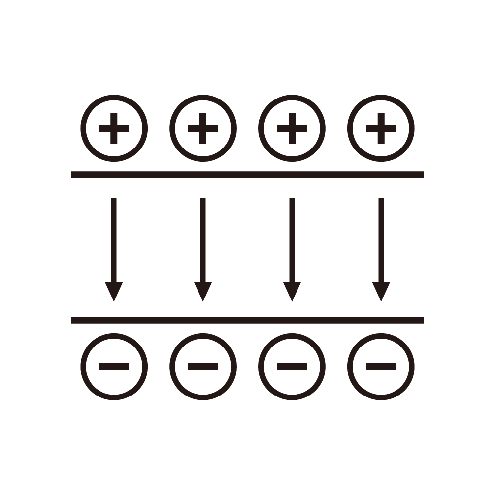
The primary advantage of low-voltage excitation is the shallow penetration depth of the primary electrons. At 1 kV, the interaction volume in a semiconductor might be only a few tens of nanometers deep, compared to a micrometer or more at 20 kV. This allows us to probe near-surface regions, thin films, and nanostructures with minimal substrate interference. However, achieving this requires that the electron beam be focused to a fine probe despite the strong chromatic aberration of the objective lens at low energies. The chromatic aberration coefficient of a magnetic lens is significant, meaning that any spread in the electron beams energy will be converted into a spread in the focus, blurring the probe. The energy spread of the beam comes from two sources: the inherent energy spread of the electron source, typically a Schottky or cold field emitter, and the ripple and drift of the high-voltage power supply that accelerates the electrons. For high-resolution imaging and analysis, the total energy spread must be kept to a minimum. At low voltages, the relative contribution of the power supply instability becomes much more significant. A 1 volt ripple on a 20 kV beam is a 0.005% perturbation, but the same 1 volt ripple on a 1 kV beam is a 0.1% perturbation, a twenty-fold increase in its effect on the focus. Therefore, the high-voltage supply for low-voltage cathodoluminescence must achieve stabilities and ripple levels that were once the exclusive domain of the most precise laboratory instruments, often in the parts-per-million range.
The generation of the cathodoluminescence signal itself is also intimately linked to the beam energy. The efficiency of light production in many materials is a strong function of the electron energy. As the beam energy is lowered, the probability of non-radiative recombination at the surface can increase, reducing the luminescence yield. The operator must be able to finely adjust the landing energy of the electrons to optimize the signal. This requires a high-voltage supply that is not only stable but also continuously and precisely adjustable over a wide range, from perhaps 500 V to 30 kV, with excellent linearity and repeatability. The ability to perform spectroscopy, where the cathodoluminescence signal is measured as a function of beam energy, demands that the voltage be settable with high resolution and that the value be accurately known. This is often achieved through a precision resistive voltage divider and a high-resolution digital-to-analog converter controlling the supply's reference. The divider itself must have an exceptionally low temperature coefficient to ensure that the reported voltage matches the actual accelerating potential.
Furthermore, the beam blanking and deflection systems, which are used to control the dwell time of the beam on the sample for panchromatic or monochromatic imaging, must operate in perfect synchrony with the high-voltage supply. For hyperspectral imaging, where a full spectrum is acquired at each pixel, the beam is often parked and then moved in a step-and-repeat fashion. The high-voltage must be absolutely stable during the entire acquisition, which can last for minutes or even hours. Any drift in the accelerating potential will cause a corresponding shift in the beam position on the sample, blurring the spectral map. In some advanced techniques, the beam energy is intentionally varied during the scan to perform depth-profiling cathodoluminescence. This requires a high-voltage supply capable of making rapid, precise steps in voltage, settling to a high accuracy without overshoot, all while maintaining the beam focus through a dynamic correction of the lens currents. In my years of consulting on microscope development, I have seen the high-voltage power supply evolve from a simple, static source into a dynamic, programmable element of the electron-optical column. For low-voltage cathodoluminescence, it is no longer just an accelerator; it is a precision instrument that, in concert with the electron optics, allows us to gently and precisely probe the optical secrets hidden in the topmost layers of our materials.