


Spatiotemporal Dynamics Study of Cell Membrane Electroporation Induced by High Voltage Pulsed Electric Field
High voltage pulsed electric fields can induce electroporation in cell membranes. Electroporation creates temporary pores that enable molecular transport across the membrane. The spatiotemporal dynamics of pore formation and evolution affect the outcome. Understanding these dynamics enables optimization of electroporation applications.
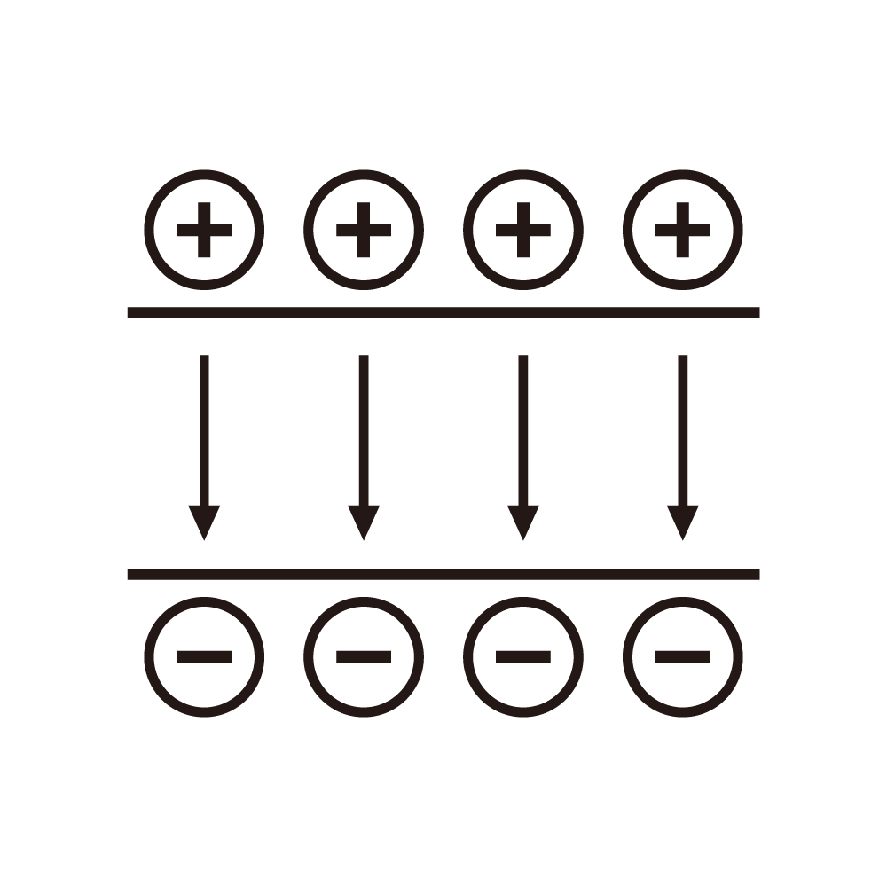
Electroporation fundamentals involve membrane permeabilization. The cell membrane is a lipid bilayer. The bilayer has inherent electrical properties. An applied electric field creates transmembrane potential. When the potential exceeds a threshold, pores form. The pores allow molecular transport.
Applications of electroporation include several fields. Drug delivery uses electroporation for cellular uptake. Gene transfection uses electroporation for DNA delivery. Food processing uses electroporation for extraction. Cancer treatment uses electroporation for therapy. Each application has specific requirements.
Transmembrane potential dynamics are critical. The applied field induces the potential. The potential depends on cell size and shape. The potential varies around the cell circumference. The potential reaches maximum at the poles. The dynamics determine the pore initiation.
Pore formation dynamics involve nucleation and growth. Pores nucleate when the threshold is exceeded. The nucleation occurs preferentially at the poles. The pores grow under continued field application. The pore size distribution evolves with time. The dynamics affect the molecular transport.
Spatial distribution of pores is non-uniform. Pores concentrate at the cell poles. The distribution depends on the field geometry. The distribution affects the transport pathways. The spatial pattern influences the cell response. The distribution must be characterized.
Temporal evolution of pores is complex. Pores form within nanoseconds of field application. The pores expand during the pulse. The pores may reseal after the pulse. The resealing time depends on the conditions. The temporal dynamics affect the outcome.
Pulse parameters affect the dynamics. The pulse amplitude determines the field strength. The pulse duration affects the pore growth. The pulse shape affects the charging dynamics. The parameters must be optimized. The optimization must consider the dynamics.
Cell factors affect the electroporation. Cell size affects the induced potential. Cell shape affects the field distribution. Membrane composition affects the threshold. The cell factors must be considered. The factors affect the optimization.
Medium factors influence the process. Conductivity affects the field distribution. Temperature affects the membrane properties. The medium factors must be controlled. The factors affect the reproducibility. The factors must be documented.
Molecular transport through pores is the goal. The transport depends on pore size. The transport depends on molecular size. The transport depends on concentration gradient. The transport must be efficient. The transport must be controlled.
Cell viability after electroporation is important. Excessive poring can cause cell death. The resealing must be complete. The viability must be maintained for applications. The viability depends on the parameters. The viability must be optimized.
Imaging techniques enable observation of dynamics. Fluorescence microscopy shows the membrane changes. Electron microscopy shows the pore structures. The imaging must have adequate resolution. The imaging must have adequate speed. The imaging data support the understanding.
Modeling of electroporation dynamics enables prediction. Continuum models describe the membrane behavior. Molecular dynamics simulate the pore formation. The models must be validated. The models enable optimization. The modeling supports the experimental work.