


Regulation Research of Microbial Cell Membrane Pore Size by High Voltage Pulsed Electric Field
The application of high voltage pulsed electric fields to microbial cells induces transmembrane potentials that can exceed the dielectric breakdown threshold of the lipid bilayer membrane, creating pores that compromise membrane integrity. This electroporation phenomenon has significant applications in biotechnology for genetic transformation, food preservation, and medical treatments. Research into the relationship between pulsed electric field parameters and the resulting pore characteristics enables optimization of electroporation processes for specific applications requiring particular pore sizes and resealing behaviors.
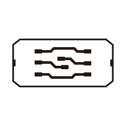
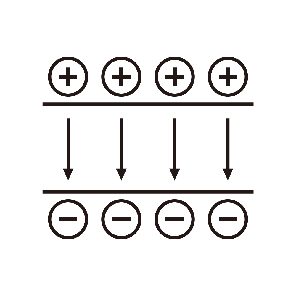
The cell membrane functions as a dielectric barrier separating the intracellular and extracellular environments, maintaining concentration gradients essential for cellular function. When an external electric field is applied, charges accumulate at the membrane surfaces, creating a transmembrane potential that adds to the natural resting potential. For spherical cells, the induced transmembrane potential depends on the cell radius, the applied field strength, and the angular position relative to the field direction. The maximum potential occurs at the poles aligned with the field direction, where electroporation initiates when the total transmembrane potential exceeds the critical threshold typically around one volt.
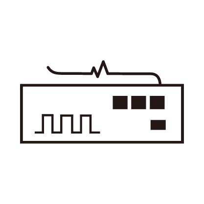
High voltage pulsed electric field systems for electroporation research must provide precise control of pulse parameters including amplitude, duration, number, and repetition rate. The pulse amplitude determines the electric field strength in the treatment chamber, which directly affects the induced transmembrane potential. Pulse duration influences the kinetics of pore formation and expansion, with longer pulses allowing more time for pores to grow. The number of pulses affects the cumulative membrane damage and the probability of achieving irreversible electroporation. The interval between pulses allows partial membrane resealing, affecting the state of the membrane for subsequent pulses.
Pore formation begins with the creation of hydrophilic pores when the transmembrane potential exceeds the critical value. These initial pores are small, on the order of nanometers, and may reseal spontaneously if the electric field is removed. Continued application of the field drives pore expansion, with larger pores allowing passage of larger molecules. The pore size distribution evolves during the pulse application, with the distribution characteristics depending on the pulse parameters and membrane properties. Understanding this evolution enables selection of pulse parameters that produce pore sizes appropriate for the intended application.
Molecular transport through electroporation pores depends on the pore size relative to the molecular dimensions. Small molecules such as ions and small metabolites can pass through even the smallest hydrophilic pores, causing immediate changes in intracellular concentrations. Larger molecules such as proteins and nucleic acids require larger pores for passage, with the transport efficiency depending on the number and size of pores large enough to accommodate the molecule. Applications requiring introduction of large molecules into cells must achieve sufficient pore expansion without causing irreversible damage to cellular functions.
The resealing dynamics after pulse application determine whether electroporation is reversible or irreversible. Reversible electroporation allows cells to survive and recover after temporary membrane permeabilization, enabling applications such as genetic transformation where cell viability is required. Irreversible electroporation causes permanent membrane damage leading to cell death, applicable for microbial inactivation in food preservation or tissue ablation in medical treatments. The transition between reversible and irreversible regimes depends on pulse parameters, with higher field strengths, longer durations, and greater pulse numbers favoring irreversible outcomes.
Experimental characterization of pore size employs various indirect and direct measurement approaches. Molecular uptake studies measure the intracellular accumulation of molecules of different sizes, inferring pore characteristics from the size dependence of uptake. Conductivity measurements of cell suspensions during and after pulse application reflect the membrane permeability changes associated with pore formation. Advanced microscopy techniques can visualize pore formation in model membrane systems, providing direct observation of pore dynamics. These characterization methods provide data for validating theoretical models of electroporation.
Mathematical models of electroporation describe the pore formation, expansion, and resealing processes in terms of membrane physical properties and applied field parameters. The models typically treat pores as having a distribution of sizes that evolves according to rate equations describing pore creation, expansion, and destruction. The pore creation rate depends exponentially on the transmembrane potential, reflecting the electric field contribution to the energy barrier for pore formation. Pore expansion occurs when the electric field energy associated with the pore exceeds the pore edge energy that tends to minimize pore size. These models enable prediction of pore characteristics for arbitrary pulse waveforms.
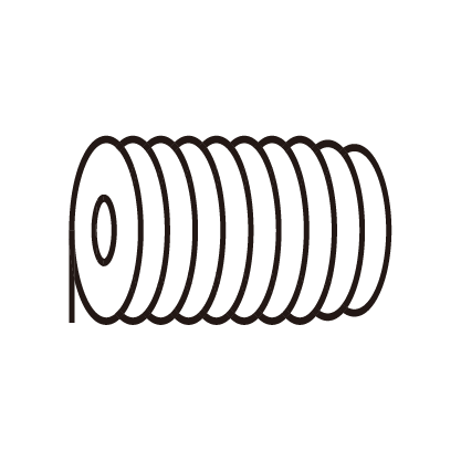
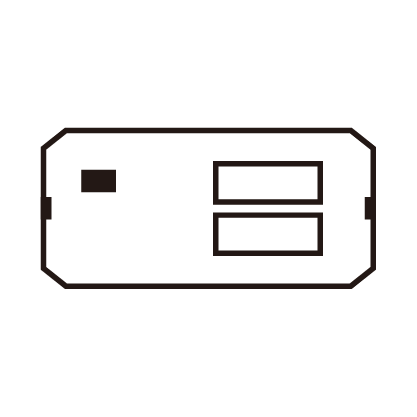
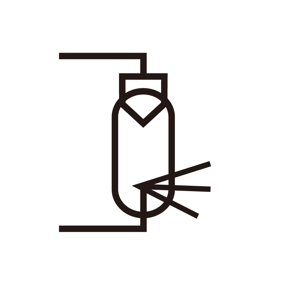
Treatment chamber design affects the uniformity of electric field exposure across the cell population. Parallel plate electrodes produce uniform fields in the gap region, but edge effects create field nonuniformities near the electrode boundaries. Coaxial and cylindrical electrode geometries produce radially varying fields that expose cells at different radial positions to different field strengths. Flow through treatment chambers ensure that all cells experience similar cumulative exposure by moving cells through the field region. Chamber design optimization considers the tradeoffs between field uniformity, treatment capacity, and electrode configuration complexity.
Temperature effects accompany electroporation due to Joule heating from current flow in the conductive medium. The temperature rise depends on the field strength squared, the medium conductivity, and the pulse duration. Excessive heating can cause thermal damage to cells in addition to the electroporation effects, complicating the interpretation of treatment outcomes. Temperature monitoring and control during pulse application ensure that thermal effects remain within acceptable bounds or are properly accounted for in the analysis of electroporation results.
Applications of pore size regulation research span multiple fields with different requirements for electroporation outcomes. Genetic transformation applications require reversible electroporation with pores large enough for nucleic acid entry but small enough to maintain cell viability. Food preservation applications exploit irreversible electroporation for microbial inactivation, with optimization focusing on achieving complete inactivation with minimum energy consumption. Medical applications such as electrochemotherapy combine reversible electroporation with drug administration to enhance drug uptake in target tissues. Each application drives specific optimization targets for pulse parameters and pore characteristics.